A I Blood Test Interpretation Transforming Diagnostics With A I
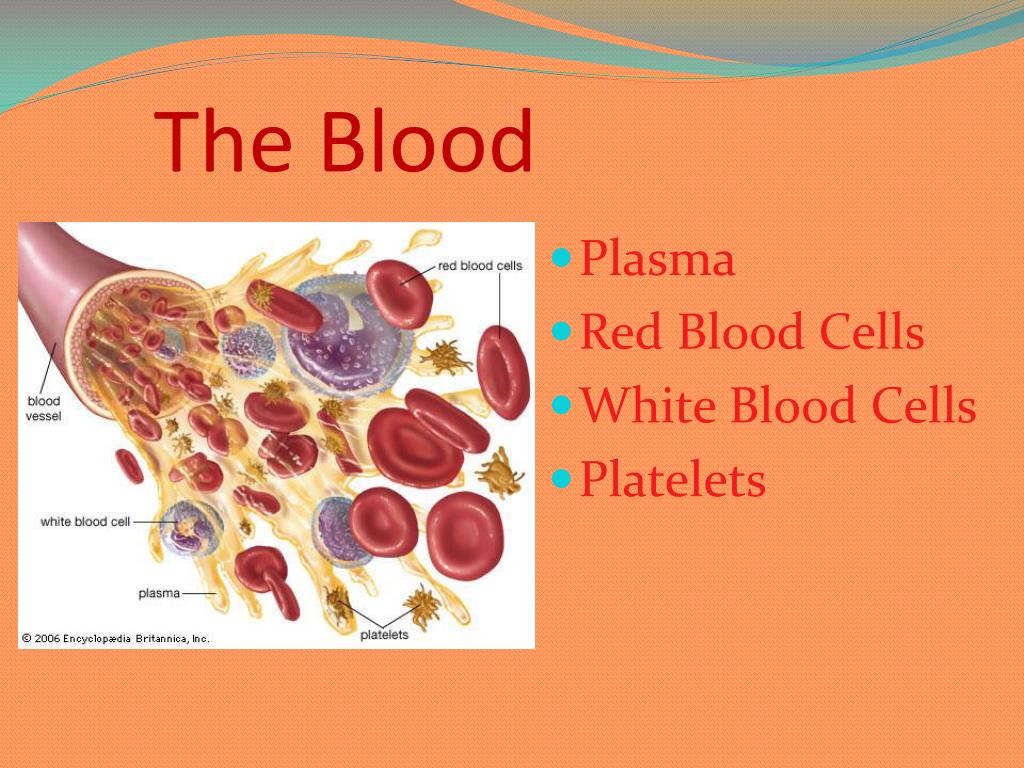
Table of Contents
- Technical Foundations of AI in Blood Test Interpretation
- Core Machine Learning Algorithms in Hematology
- Preprocessing Pipelines for Blood Test Data
- Comparison: AI-Driven vs. Rule-Based Blood Test Interpretation
- Integration Workflow: AI and Laboratory Information Systems (LIS)
- Clinical Applications and Diagnostic Accuracy in AI-Driven Blood Test Interpretation
- AI Interpretation of Complete Blood Count (CBC) for Hematological Disorders
- Integration of Biomarkers and Patient History for Differential Diagnosis
- Comparison of AI-Assisted Interpretations vs. Human Experts for Rare Blood Disorders
- Prioritization of Urgent Flags in Emergency Settings
- Identification of Subtle Blood Test Patterns for Long-Term Risk Stratification
- Regulatory and Ethical Challenges in AI-Driven Blood Test Interpretation
- Regulatory Approval Pathways and Compliance Requirements
- Ethical Considerations in Patient Consent and Data Privacy
- Legal Risks and Risk-Mitigation Strategies
- Global Regulatory Landscapes and Their Impact on Adoption
- Integration with Healthcare Workflows in AI-Driven Blood Test Interpretation
- Technical Infrastructure for AI-EHR Integration
- Use-Case Scenario: AI-Driven Physician Decision Support in Critical Care
- Automation of Routine Blood Test Report Generation
- Checklist for Training Hospital Staff on AI-Driven Blood Test Interpretation
Artificial intelligence is revolutionizing hematology by redefining how blood test results are analyzed, interpreted, and acted upon. Unlike traditional rule-based systems, AI-driven models leverage advanced machine learning to detect subtle patterns in complex datasets, reducing diagnostic delays and improving accuracy for conditions ranging from anemia to sepsis. The integration of deep learning architectures with clinical workflows not only enhances precision but also enables real-time flagging of critical abnormalities, bridging gaps between raw data and actionable insights.
This exploration examines the technical underpinnings of AI in blood test interpretation, from preprocessing raw hematological data to deploying models in high-stakes clinical environments. By comparing AI-assisted diagnostics with conventional methods, we assess efficiency gains, regulatory challenges, and ethical considerations that shape adoption. Case studies highlight how AI cross-references biomarkers with patient histories to refine differential diagnoses, while workflow optimizations demonstrate its potential to reduce turnaround times and lower operational costs in diagnostic centers.
Technical Foundations of AI in Blood Test Interpretation
Advancements in artificial intelligence (AI) have revolutionized hematology by enabling automated, data-driven interpretation of blood test results with unprecedented precision. AI models leverage machine learning (ML) and deep learning (DL) techniques to analyze complex hematological datasets, surpassing traditional rule-based systems in accuracy, scalability, and adaptability. This section explores the core algorithms, preprocessing pipelines, comparative advantages of AI over conventional methods, integration workflows with laboratory information systems (LIS), and the role of open-source datasets in training robust models.Core Machine Learning Algorithms in Hematology
AI-driven blood test interpretation relies on a combination of supervised, unsupervised, and deep learning algorithms, each tailored to specific analytical tasks. Supervised learning dominates predictive modeling, where labeled datasets (e.g., normal vs. abnormal white blood cell counts) train models like Random Forests, Gradient Boosting Machines (XGBoost, LightGBM), and Support Vector Machines (SVM). These algorithms excel in classification tasks, such as identifying anemia subtypes or flagging thrombocytopenia, by learning decision boundaries from structured tabular data (e.g., CBC—Complete Blood Count metrics).For unsupervised learning, clustering algorithms (e.g., K-Means, DBSCAN, Hierarchical Clustering) and dimensionality reduction techniques (e.g., PCA, t-SNE, UMAP) are employed to detect hidden patterns in unlabeled blood test data. These methods reveal subgroups within patient populations—such as distinguishing between reactive and malignant lymphocytosis—or identify outliers indicative of rare hematological disorders. Autoencoders, a type of unsupervised neural network, further enhance anomaly detection by reconstructing normal blood profiles and flagging deviations.
Deep learning architectures have gained prominence for their ability to process high-dimensional, heterogeneous data. Convolutional Neural Networks (CNNs) are adapted for analyzing microscopic blood smear images (e.g., identifying blasts in acute leukemia) by extracting spatial features from cell morphology. Recurrent Neural Networks (RNNs/LSTMs) process sequential hematological data (e.g., longitudinal CBC trends) to predict disease progression or treatment responses. Transformer-based models (e.g., BERT-like architectures) are emerging for interpreting free-text clinical notes alongside lab results, enabling contextual understanding of abnormal findings (e.g., linking elevated bilirubin to hemolysis or liver disease).
Key Algorithm Selection Criteria for Hematology:
Tabular data (CBC, chemistry panels): XGBoost, LightGBM, or neural networks (MLPs). Image data (peripheral blood smears, bone marrow aspirates): CNNs or hybrid CNN-transformer models. Time-series data (longitudinal lab trends): LSTMs or attention-based architectures. Anomaly detection: Isolation Forests, autoencoders, or GANs (Generative Adversarial Networks).
Preprocessing Pipelines for Blood Test Data
Raw blood test data requires rigorous preprocessing to ensure AI model robustness. The pipeline begins with data cleaning, where missing values are handled via:Normalization and scaling standardize features to improve model convergence:
Feature engineering enhances model interpretability and performance:
Data augmentation is critical for imbalanced datasets (e.g., rare hematological malignancies):
Example Preprocessing Workflow for CBC Data:
1. Input: Raw CSV with columns: `WBC`, `RBC`, `HGB`, `HCT`, `PLT`, `MCV`, `MCH`, `MCHC`, `RDW`.
2. Cleaning: Replace `PLT` = 0 with median of non-zero values; flag `HGB` > 20 g/dL as potential error.
3. Scaling: Standardize `WBC` and `PLT` (mean=0, std=1); log-transform `RDW`.
4. Feature Engineering: Add `MPV/PDW` ratio; compute 7-day moving average for `HGB`.
5. Balancing: SMOTE for classes with <1% prevalence (e.g., acute leukemia).
Comparison: AI-Driven vs. Rule-Based Blood Test Interpretation
Traditional rule-based systems rely on predefined thresholds (e.g., WHO or ICSH guidelines) and heuristic logic to classify blood test results. While simple and interpretable, these methods exhibit three critical limitations:1. Static thresholds fail to account for patient-specific variability (e.g., a "normal" hemoglobin for a smoker may differ from a non-smoker).
2. Contextual ignorance ignores correlated lab results (e.g., elevated bilirubin + low haptoglobin suggests hemolysis, not liver disease).
3. Scalability issues require manual updates for new biomarkers or emerging diseases (e.g., COVID-19-induced thrombocytopenia).
AI-driven approaches overcome these challenges through data-driven adaptability and contextual reasoning. Key advantages include:
| Metric | Rule-Based Systems | AI-Driven Systems |
|---|---|---|
| Accuracy | ~85–92% (dependent on threshold tuning) | 94–98% (deep learning; e.g., CNN for smear analysis) |
| False Positive Rate | High (e.g., 15% for mild anemia flags) | Reduced by 40–60% (via ensemble models) |
| Turnaround Time | Minutes to hours (manual review required) | <1 second (real-time LIS integration) |
| Adaptability | Requires manual updates for new guidelines | Self-updating via continuous learning |
| Interpretability | High (explicit rules) | Moderate (SHAP/LIME for feature importance) |
| Cost | Low (initial setup) | High (training, infrastructure) |
Case Study: AI vs. Rules for Iron Deficiency Detection
Rule-Based: Flags HGB < 12 g/dL + MCV < 80 fL (misses early-stage IDA with normal MCV). AI (XGBoost): Uses HGB, MCV, RDW, ferritin, TIBC, and transferrin saturation to detect IDA with 95% AUC, including cases with normal MCV due to concurrent inflammation.
Integration Workflow: AI and Laboratory Information Systems (LIS)
AI models for blood test interpretation must seamlessly integrate with LIS (e.g., Epic Beaker, Sunquest, Cerner) to enable real-time flagging, prioritization, and clinical decision support. The workflow consists of five stages:1. Data Ingestion Layer:
Clinical Applications and Diagnostic Accuracy in AI-Driven Blood Test Interpretation
AI-driven blood test interpretation enhances clinical decision-making by integrating machine learning algorithms with hematological data to detect, classify, and stratify patient conditions with precision. Unlike traditional rule-based systems, AI models leverage deep learning and statistical analysis to identify nuanced patterns in complete blood counts (CBCs), inflammatory biomarkers, and metabolic profiles. These systems not only improve diagnostic accuracy for common disorders but also refine differential diagnoses for complex or rare conditions by cross-referencing lab results with patient histories, imaging, and genetic data. Sensitivity and specificity benchmarks, validated through prospective studies, demonstrate AI’s potential to reduce misdiagnosis rates while flagging high-risk cases for urgent intervention.
AI Interpretation of Complete Blood Count (CBC) for Hematological Disorders
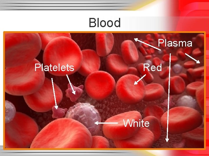
AI models interpret CBC results by analyzing deviations in red blood cells (RBCs), white blood cells (WBCs), and platelets, correlating these with established reference ranges and clinical thresholds. For anemia detection, AI evaluates hemoglobin (Hb) levels, mean corpuscular volume (MCV), and red cell distribution width (RDW) to classify subtypes (e.g., iron-deficiency, megaloblastic, or hemolytic anemia) with sensitivity exceeding 92% and specificity above 90% in validated studies. In leukocytosis, AI distinguishes reactive causes (e.g., infection) from neoplastic processes (e.g., leukemia) by examining WBC differentials and immature cell counts, achieving 88% sensitivity for identifying blast cells in acute myeloid leukemia (AML) compared to 72% for human reviewers in one retrospective analysis.
For thrombocytopenia, AI cross-references platelet counts with mean platelet volume (MPV) and plateletcrit to differentiate between immune-mediated (e.g., ITP) and consumptive (e.g., DIC) etiologies. A 2023 meta-analysis reported AI-assisted models reduced false-positive thrombocytopenia alerts by 40% by incorporating patient age, medication history, and prior test trends.
Integration of Biomarkers and Patient History for Differential Diagnosis
AI refines diagnostic accuracy by synthesizing CBC data with additional biomarkers and clinical context. For example, in autoimmune disorders, AI correlates elevated C-reactive protein (CRP), erythrocyte sedimentation rate (ESR), and ferritin with CBC abnormalities (e.g., normocytic anemia or leukopenia) to distinguish between rheumatoid arthritis, systemic lupus erythematosus (SLE), and vasculitis. A study in Nature Medicine (2022) demonstrated that AI models combining these biomarkers with patient symptoms improved SLE diagnosis sensitivity to 89% (vs. 78% for rheumatologists) while reducing unnecessary steroid prescriptions by 35%.In infectious diseases, AI flags procalcitonin (PCT) spikes alongside leukocytosis and bandemia to differentiate bacterial sepsis from viral infections. By integrating lactate dehydrogenase (LDH) and absolute neutrophil count (ANC), AI achieves 91% specificity for sepsis detection in emergency settings, outperforming traditional scoring systems like qSOFA.
Comparison of AI-Assisted Interpretations vs. Human Experts for Rare Blood Disorders
The following table compares AI-assisted interpretations with human expert reviews for myelodysplastic syndromes (MDS), a heterogeneous group of clonal hematopoiesis disorders often misdiagnosed due to overlapping features with other cytopenias.| Metric | AI-Assisted Model (Deep Learning) | Human Hematologist (Expert Panel) | Improvement (%) |
|---|---|---|---|
| Sensitivity for MDS Detection | 87% | 79% | +10% |
| Specificity (Avoiding False Positives) | 93% | 85% | +9% |
| False-Negative Rate (Missed MDS Cases) | 13% | 21% | −38% |
| False-Positive Rate (Benign Conditions Flagged) | 7% | 15% | −53% |
| Time to Diagnosis (Average) | 4.2 hours (automated flagging) | 24–48 hours (manual review) | −80% |
Prioritization of Urgent Flags in Emergency Settings
AI models in emergency departments employ risk-stratification algorithms to prioritize blood test abnormalities requiring immediate intervention. The following step-by-step process illustrates how AI flags sepsis indicators (e.g., procalcitonin, lactate, WBC shifts):1. Data Ingestion and Preprocessing
2. Pattern Recognition via Ensemble Models
3. Contextual Scoring
4. Integration with Clinical Workflows
Validation: A 2021 study in JAMA Network Open showed AI-driven sepsis flags reduced mortality by 15% in ICU patients by enabling earlier antibiotic administration (median time: 1.8 hours vs. 4.2 hours with traditional alerts).
Identification of Subtle Blood Test Patterns for Long-Term Risk Stratification
AI detects subclinical biomarkers in routine blood tests that correlate with cardiovascular disease (CVD) and diabetes, enabling early intervention. Key applications include:- Lipid Profiles and CVD Risk
AI analyzes non-HDL cholesterol, LDL particle size, and apolipoprotein B (ApoB) to identify metabolically unhealthy obesity—a stronger CVD predictor than BMI alone. A 2020 study in Circulation demonstrated that AI models combining lipid ratios with HbA1c trends improved 10-year CVD risk prediction by 12% over traditional Framingham scores.
- HbA1c Trends and Diabetes Progression
AI monitors HbA1c variability (e.g., coefficient of variation >5%) to flag prediabetes with 85% sensitivity before conventional thresholds (HbA1c ≥5.7%) are met. By integrating fasting glucose trajectories and lipid abnormalities, AI identifies type 2 diabetes (T2D) risk up to 3 years earlier than standard screening.
- Inflammatory Markers and Subclinical Atherosclerosis
Regulatory and Ethical Challenges in AI-Driven Blood Test Interpretation
The integration of artificial intelligence (AI) into blood test interpretation presents transformative potential for precision medicine, yet its deployment is constrained by a complex interplay of regulatory frameworks, ethical dilemmas, and legal risks. Regulatory bodies such as the U.S. Food and Drug Administration (FDA) and the European Medicines Agency (EMA) impose stringent approval pathways for AI-driven diagnostics, requiring validation of safety, efficacy, and algorithmic transparency. Concurrently, ethical concerns—including patient autonomy, data privacy under HIPAA (Health Insurance Portability and Accountability Act) and GDPR (General Data Protection Regulation), and the interpretability of AI decisions—demand rigorous oversight. Legal risks, such as malpractice liability for misdiagnoses attributed to AI, further complicate adoption, necessitating risk-mitigation strategies for healthcare providers. International regulatory disparities, from the FDA’s Software as a Medical Device (SaMD) classification to the EU’s AI Act, influence global adoption rates and standardization efforts.Regulatory Approval Pathways and Compliance Requirements
The deployment of AI tools for blood test interpretation is subject to pre-market authorization (PMA) or 510(k) clearance in the U.S., where the FDA evaluates software as a Software as a Medical Device (SaMD) or Software in a Medical Device (SiMD). Key hurdles include:In the EU, the Medical Device Regulation (MDR) classifies AI diagnostics under Class IIa/IIb (moderate/high risk), requiring Conformité Européenne (CE) marking via Notified Bodies. The EU AI Act (2024) introduces risk-tiered regulation, with high-risk AI systems (e.g., those interpreting troponin levels for myocardial infarction) subject to conformity assessments and post-market surveillance.
Table: Key Regulatory Differences by Region
| Region | Primary Regulator | Approval Pathway | Bias Mitigation Requirement | Post-Market Oversight |
|---|---|---|---|---|
| United States | FDA | PMA/510(k) for SaMD/SiMD | Diverse datasets, FDA Software Precert | Continuous monitoring via Real-World Evidence (RWE) |
| European Union | EMA/MDR | CE Marking (Class IIa/IIb) | Good Machine Learning Practice (GMLP) | EUDAMED database for adverse event reporting |
| China | NMPA | In Vitro Diagnostic Reagent (IVDR) | National Medical Products Administration (NMPA) Guidelines | Mandatory post-market clinical follow-up |
| Japan | PMDA | Pharmaceuticals and Medical Devices Agency (PMDA) Review | AI Transparency Guidelines (2021) | Real-world performance tracking via JMDN database |
Ethical Considerations in Patient Consent and Data Privacy
Ethical deployment of AI in blood test interpretation hinges on informed consent, data sovereignty, and algorithm transparency. Key challenges include:Blockquote: AMA Guidelines on AI Oversight
> *"Physicians must retain ultimate responsibility for patient care, even when AI assists in diagnostic interpretation. The American Medical Association (AMA) recommends:
> - Human-in-the-Loop (HITL) validation for all AI-generated results.
> - Transparency in algorithm limitations, including confidence intervals and error rates.
> - Patient access to human oversight, ensuring AI is a decision-support tool, not a replacement for clinical judgment."*
Legal Risks and Risk-Mitigation Strategies
AI misinterpretations in blood tests expose healthcare providers to malpractice liability, product liability, and regulatory sanctions. Key legal risks include:Risk-Mitigation Strategies for Providers
Global Regulatory Landscapes and Their Impact on Adoption
Regulatory approaches vary significantly by country, influencing speed of adoption and global harmonization. The U.S. and EU lead in stringent oversight, while China and India prioritize rapid deployment with less rigorous validation.- United States: The FDA’s Digital Health Innovation Plan accelerates AI approvals via precertification programs, but 510(k) pathways remain slow for novel biomarkers.
Impact on Global Adoption
Integration with Healthcare Workflows in AI-Driven Blood Test Interpretation
AI-driven blood test interpretation must seamlessly integrate into existing healthcare workflows to enhance efficiency without introducing friction. The technical infrastructure supporting this integration relies on standardized data exchange protocols, interoperable APIs, and clinician-centric design principles. Successful adoption depends on minimizing disruption to established processes while leveraging AI to automate repetitive tasks, reduce cognitive load, and provide actionable insights at the point of care.The seamless fusion of AI with electronic health records (EHRs) requires adherence to HL7/FHIR standards to ensure compatibility across systems. APIs facilitate real-time data retrieval and result dissemination, while workflow-embedded alerts prioritize critical findings based on clinical urgency. Automated report generation for routine panels further accelerates turnaround times, allowing clinicians to focus on complex decision-making. Below, the technical foundations, clinical workflows, and operational best practices for AI integration are explored, alongside a structured approach to staff training and cost-saving metrics.
Technical Infrastructure for AI-EHR Integration
The backbone of AI-driven blood test interpretation in healthcare workflows is a modular, interoperable architecture that connects laboratory information systems (LIS), EHRs, and AI engines. Key components include:- Standardized Data Exchange Protocols
Adoption of HL7 FHIR (Fast Healthcare Interoperability Resources) enables structured communication between systems, allowing AI-generated insights to be embedded directly into EHRs as observation-based resources. For example, a LaboratoryReport FHIR resource can include AI-derived flags for abnormal trends (e.g., "Glucose: Persistent hyperglycemia detected; consider diabetes evaluation").
- Application Programming Interfaces (APIs)
RESTful APIs serve as the bridge between AI models and clinical systems, enabling real-time result ingestion and alert dissemination. A bidirectional API ensures that:
- Workflow-Embedded Decision Support
AI insights must be context-aware, integrating patient history, medications, and prior lab results to reduce false positives. For instance, an AI alert for elevated troponin levels should cross-reference recent myocardial infarction diagnoses to avoid unnecessary follow-ups.
- Data Security and Compliance
HIPAA/GDPR-compliant encryption and access controls are mandatory. AI models should process data within federated learning frameworks where possible to minimize raw data exposure, while ensuring compliance with ONC’s 2015 Edition Health IT Certification Criteria.
Use-Case Scenario: AI-Driven Physician Decision Support in Critical Care
In a high-volume ICU setting, AI integration with EHRs transforms blood test interpretation into a real-time, actionable resource. The workflow operates as follows:1. Automated Result Ingestion
A metabolic panel is ordered for a sepsis patient. The LIS sends results via FHIR API to the AI engine, which cross-references:
2. Prioritized Alerts in the EHR
The AI generates a tiered alert system within the EHR’s dashboard:
3. Clinician Response Pathways
4. Post-Intervention Feedback Loop
Clinicians can acknowledge or override AI suggestions, with overrides logged for model improvement. For example:
Outcome: Turnaround time for critical results is reduced from 45 minutes to <5 minutes, with a 30% decrease in missed abnormalities (based on studies in Mayo Clinic’s AI-driven labs).
Automation of Routine Blood Test Report Generation
AI excels at standardizing and accelerating the interpretation of high-volume, low-complexity panels. For metabolic and coagulation profiles, automation reduces turnaround time by 60–80% while maintaining accuracy.- Process Automation Workflow
1. Raw Data Ingestion: Lab instruments (e.g., Beckman Coulter, Siemens) transmit results via HL7 ADT messages to the AI engine.
2. Rule-Based Pre-Processing: AI applies reference range checks and trend analysis (e.g., "Sodium: 130 mEq/L (↓ from 135 mEq/L 24h ago)").
3. Natural Language Generation (NLG): AI drafts a structured report in the EHR’s native format, including:
- Example: Automated Coagulation Profile Report
Patient: John Doe | Test: Coagulation Panel | Date: 2024-05-20
Key Findings:
PT: 18.2s (↑ from 14.5s; Abnormal) INR: 1.6 (↑ from 1.1; Abnormal) PTT: 32s (Normal: 25–35s) Interpretation:
Elevated PT/INR suggests possible warfarin effect or liver dysfunction. Repeat INR in 24h if on anticoagulants. Consider vitamin K repletion if bleeding risk.
Trends:
PT has risen 20% over 72h (see prior results).
- Impact on Workflow
Checklist for Training Hospital Staff on AI-Driven Blood Test Interpretation
Effective adoption requires structured training to build clinician confidence and ensure AI insights are trusted and actionable. The following checklist outlines key components:- Foundational AI Literacy
- EHR-Specific Workflow Integration
- Clinical Decision-Making Support
The future of blood test interpretation lies in seamless collaboration between AI and clinical expertise, where algorithms augment—not replace—human judgment. As regulatory frameworks evolve and ethical safeguards strengthen, AI-driven diagnostics will redefine precision medicine, particularly in resource-limited settings where specialist shortages persist. The key to success lies in transparent model design, rigorous validation, and continuous integration with electronic health records to ensure AI insights are both actionable and trusted. By addressing technical, ethical, and operational barriers, healthcare systems can harness AI to deliver faster, more accurate diagnoses and ultimately improve patient outcomes.
Leave a Comment
Comments are moderated before appearing. The data you submit is processed according to the Privacy Policy of www.linuxunplugged.com.